
Thermal Camera
Imaging technique of cancer and muscle diseases, breast cancer and treatment impressions by detecting body temperature differences with an invisible (infaruge) high-resolution thermal camera.
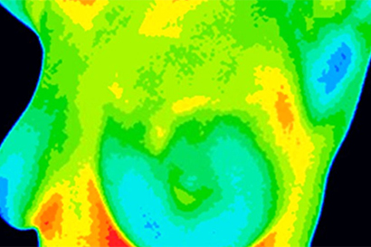
DITI (Breast Thermalgraphy): The device does not emit any rays to the body, it is the process of detecting the thermal - heat rays (infrared) emitted from the body with the help of a special camera. Since it is highly sensitive to temperature, it can detect temperature changes in breast tissue even at a very early stage.
However, it cannot show cell structures. For this reason, ultrasound is used as an auxiliary technique to evaluate the risk and potential for change of masses that can be detected by other techniques such as mammography. We mentioned that cancer cells need more nutrition and blood. This increase in blood flow to the tissue also creates a similar increase in blood flow and temperature in the projections of the tissues on the skin. The device detects the increase in temperature on the skin due to the reaction of the body, rather than the direct heat of the organ.
Benign structures generally do not cause any symptoms in Diti because they do not have a significant blood supply. Fibrocystic structures cause a slight increase in blood flow. Usually this is symmetrical as it occurs in both breasts. Precancerous structures show a greater temperature increase symmetrically. However, since no obvious structure can be detected in other techniques, the patient is closely monitored. Excessive and asymmetric temperature increases are considered highly suspicious for developing or developing structures. To understand the reason for this difference, further examinations should be performed and the patient should be followed closely.
• Healthy Breast Tracking
• Breast Diseases Follow-up
• Breast Cancer Diagnosis and Follow-up
• Monitoring the response to Neoadjuvant Chemotherapy in breast cancer
• Follow-up after breast cancer